
Page 177 - Atlas of Histology with Functional Correlations
P. 177
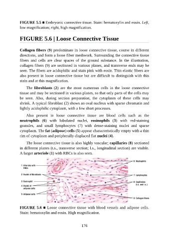
FIGURE 5.5 ■ Embryonic connective tissue. Stain: hematoxylin and eosin. Left,

               low magnification; right, high magnification.


               FIGURE 5.6 | Loose Connective Tissue


               Collagen fibers (9) predominate in loose connective tissue, course in different

               directions, and form a loose fiber meshwork. Surrounding the connective tissue
               fibers  and  cells  are  clear  spaces  of  the  ground  substance.  In  the  illustration,
               collagen fibers (9) are sectioned in various planes, and transverse ends may be
               seen. The fibers are acidophilic and stain pink with eosin. Thin elastic fibers are

               also present in loose connective tissue but are difficult to distinguish with this
               stain and at this magnification.

                   The  fibroblasts  (2)  are  the  most  numerous  cells  in  the  loose  connective
               tissue and may be sectioned in various planes, so that only parts of the cells may
               be  seen.  Also,  during  section  preparation,  the  cytoplasm  of  these  cells  may

               shrink. A typical fibroblast (2) shows an oval nucleus with sparse chromatin and
               lightly acidophilic cytoplasm, with a few short processes.

                   Also  present  in  loose  connective  tissue  are  blood  cells  such  as  the
               neutrophils  (6)  with  lobulated  nuclei,  eosinophils  (3)  with  red-staining

               granules,  and  small  lymphocytes  (7)  with  dense-staining  nuclei  and  sparse
               cytoplasm. The fat (adipose) cells (5) appear characteristically empty with a thin
               rim of cytoplasm and peripherally displaced flat nuclei (4).

                   The loose connective tissue is also highly vascular; capillaries (8) sectioned
               in different planes (t.s., transverse section; l.s., longitudinal section) are visible.

               A larger arteriole (1) with RBCs is also seen.


               FIGURE 5.6 ■ Loose connective tissue with blood vessels and adipose cells.
               Stain: hematoxylin and eosin. High magnification.


                                                          176