
Page 178 - Atlas of Histology with Functional Correlations
P. 178
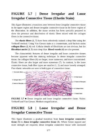
FIGURE  5.7  |  Dense  Irregular  and  Loose

               Irregular Connective Tissue (Elastin Stain)


               This figure illustrates a transition zone between loose irregular connective tissue
               in the upper region and denser irregular connective tissue in the lower region of

               the  illustration.  In  addition,  the  tissue  section  has  been  specially  prepared  to
               show  the  presence  and  distribution  of  elastic  fibers  mixed  with  the  collagen
               fibers in the connective tissue.

                   The elastic fibers (1, 7) have been selectively stained a deep blue using the
               Verhoeff method. Using Van Gieson stain as a counterstain, acid fuchsin stains

               collagen fibers (2, 6) red. Cellular details of fibroblasts are not obvious, but the
               fibroblast nuclei (3, 5) stain deep blue. Blood vessels (4) are also present.

                   The  characteristic  features  of  dense  irregular  and  loose  connective  tissues
               become  apparent  with  this  staining  technique.  In  dense  irregular  connective

               tissue, the collagen fibers (6) are larger, more numerous, and more concentrated.
               Elastic fibers are also larger and more numerous (7). In contrast, in the loose
               connective tissue, both fiber types are smaller (1, 2) and more loosely arranged.
               Fine elastic networks are seen in both types of connective tissue.


               FIGURE  5.7  ■  Dense  irregular  and  loose  irregular  connective  tissue.  Stains:

               Verhoeff and Van Gieson. Medium magnification.


               FIGURE  5.8  |  Loose  Irregular  and  Dense


               Irregular Connective Tissue


               This  figure  illustrates  a  gradual  transition  from  loose  irregular  connective
               tissue (5) to dense irregular connective tissue (1). Where firmer support and
               more  strength  are  required,  dense  irregular  connective  tissue  with  increased


                                                          177