
Page 183 - Atlas of Histology with Functional Correlations
P. 183
FUNCTIONAL CORRELATIONS 5.3 ■ Dense
Connective Tissue
Dense Irregular Connective Tissue
Dense irregular connective tissue consists primarily of collagen fibers (type
I collagen) with minimal amounts of surrounding ground substance. Except
for the fibroblasts and/or fibrocytes, other cell types in dense connective
tissue are sparse. Collagen fibers exhibit great tensile strength, and their
main function is support. In dense irregular connective tissue, collagen
fibers exhibit random orientation and are most highly concentrated in those
areas of the body where strong support is needed to resist pulling forces or
stress from different directions.
Dense Regular Connective Tissue
Dense regular connective tissue exhibits a predominance of collagen fibers
(type I collagen) and is present where great tensile strength is required,
such as in ligaments and tendons. The parallel and dense arrangements of
collagen fibers offer strong resistance to forces pulling along a single axis or
direction.
Tendons and ligaments are attached to bones and are constantly subjected
to strong pulling forces. Because of the dense arrangement of collagen fibers,
little ground substance is present, and the predominant cell types that
synthesize the collagen fibers are the fibroblasts that are located between
rows of parallel collagen fibers.
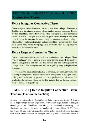
FIGURE 5.12 | Dense Regular Connective Tissue:
Tendon (Transverse Section)
A transverse section of a tendon is illustrated at a lower magnification (left side)
and a higher magnification (right side). Within each large bundle of collagen
fibers (3, 7) are fibroblasts (nuclei) (1, 8) sectioned transversely. The
fibroblasts are located between the bundles of collagen fibers (3, 7). These
fibroblasts (8) are better distinguished at the higher magnification on the right
side, which shows bundles of collagen fibers (7) and the branched shape of
182