
Page 184 - Atlas of Histology with Functional Correlations
P. 184
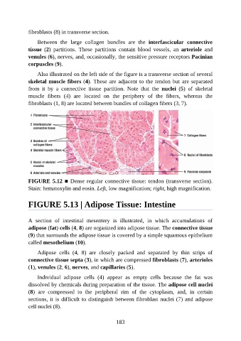
fibroblasts (8) in transverse section.

                   Between  the  large  collagen  bundles  are  the  interfascicular  connective

               tissue  (2)  partitions.  These  partitions  contain  blood  vessels,  an  arteriole  and
               venules (6), nerves, and, occasionally, the sensitive pressure receptors Pacinian
               corpuscles (9).

                   Also illustrated on the left side of the figure is a transverse section of several
               skeletal muscle fibers (4). These are adjacent to the tendon but are separated

               from  it  by  a  connective  tissue  partition.  Note  that  the  nuclei  (5)  of  skeletal
               muscle  fibers  (4)  are  located  on  the  periphery  of  the  fibers,  whereas  the
               fibroblasts (1, 8) are located between bundles of collagen fibers (3, 7).


               FIGURE 5.12 ■ Dense regular connective tissue: tendon (transverse section).

               Stain: hematoxylin and eosin. Left, low magnification; right, high magnification.


               FIGURE 5.13 | Adipose Tissue: Intestine


               A  section  of  intestinal  mesentery  is  illustrated,  in  which  accumulations  of

               adipose (fat) cells (4, 8) are organized into adipose tissue. The connective tissue
               (9) that surrounds the adipose tissue is covered by a simple squamous epithelium
               called mesothelium (10).

                   Adipose  cells  (4,  8)  are  closely  packed  and  separated  by  thin  strips  of

               connective tissue septa (3), in which are compressed fibroblasts (7), arterioles
               (1), venules (2, 6), nerves, and capillaries (5).

                   Individual  adipose  cells  (4)  appear  as  empty  cells  because  the  fat  was
               dissolved by chemicals during preparation of the tissue. The adipose cell nuclei
               (8)  are  compressed  to  the  peripheral  rim  of  the  cytoplasm,  and,  in  certain

               sections, it is difficult to distinguish between fibroblast nuclei (7) and adipose
               cell nuclei (8).


                                                          183