
Page 242 - Atlas of Histology with Functional Correlations
P. 242
bind to glycosaminoglycans and collagen fibers, providing adherence of
chondroblasts and chondrocytes to collagen fibers of the surrounding matrix.
Although hyaline cartilage contains type II collagen fibers in its matrix, in
routine histologic preparations, these collagen fibers are not seen because their
reflective index is similar to that of the surrounding ground substance.
Supplemental micrographic images are available at
www.thePoint.com/Eroschenko13e under Cartilage.
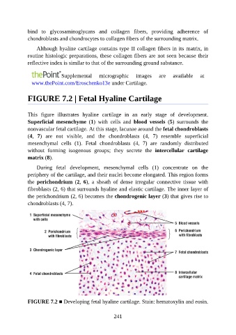
FIGURE 7.2 | Fetal Hyaline Cartilage
This figure illustrates hyaline cartilage in an early stage of development.
Superficial mesenchyme (1) with cells and blood vessels (5) surrounds the
nonvascular fetal cartilage. At this stage, lacunae around the fetal chondroblasts
(4, 7) are not visible, and the chondroblasts (4, 7) resemble superficial
mesenchymal cells (1). Fetal chondroblasts (4, 7) are randomly distributed
without forming isogenous groups; they secrete the intercellular cartilage
matrix (8).
During fetal development, mesenchymal cells (1) concentrate on the
periphery of the cartilage, and their nuclei become elongated. This region forms
the perichondrium (2, 6), a sheath of dense irregular connective tissue with
fibroblasts (2, 6) that surrounds hyaline and elastic cartilage. The inner layer of
the perichondrium (2, 6) becomes the chondrogenic layer (3) that gives rise to
chondroblasts (4, 7).
FIGURE 7.2 ■ Developing fetal hyaline cartilage. Stain: hematoxylin and eosin.
241