
Page 243 - Atlas of Histology with Functional Correlations
P. 243
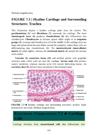
Medium magnification.


               FIGURE 7.3 | Hyaline Cartilage and Surrounding

               Structures: Trachea


               This  illustration  depicts  a  hyaline  cartilage  plate  from  the  trachea.  The

               perichondrium  (5)  with  fibroblasts  (7)  surrounds  the  cartilage.  The  inner
               chondrogenic  layer  (4)  produces  chondroblasts  (8)  that  differentiate  into
               chondrocytes.  Chondrocytes  in  lacunae  appear  either  single  or  in  isogenous

               groups (3). Lacunae and chondrocytes (3) in the middle of the cartilage plate are
               large and spherical but become flatter toward the periphery, where these cells are
               differentiating  into  chondroblasts  (8).  The  interterritorial  (intercellular)
               matrix (1) stains lighter, whereas the territorial matrix (2) around the lacunae
               stains darker.


                   Vascular  (9)  connective  tissue  (10)  and  tracheal  glands  with  grapelike
               secretory  units  called  acini  are  near  the  cartilage.  Serous  acini  (11)  produce
               watery  secretions,  whereas  mucous  acini  (12)  secrete  lubricating  mucus.  An
               excretory duct (6) delivers these secretions to the tracheal lumen.


               FIGURE  7.3  ■  Hyaline  cartilage  and  surrounding  structures:  trachea.  Stain:

               hematoxylin and eosin. Medium magnification.


                  FUNCTIONAL  CORRELATIONS  7.1  ■  Cartilage

                  Cells


                 Cartilage  develops  from  mesenchymal  cells  that  differentiate  into


                                                          242