
Page 248 - Atlas of Histology with Functional Correlations
P. 248
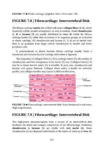
FIGURE 7.7 ■ Elastic cartilage: epiglottis. Stain: silver stain. ×80.


               FIGURE 7.8 | Fibrocartilage: Intervertebral Disk


               The fibrous cartilage matrix (5) is filled with dense collagen fibers (2, 6), which
               frequently exhibit parallel arrangement, as seen in tendons. Small chondrocytes

               (1,  4)  in  lacunae  (3)  are  usually  distributed  in  rows  (4)  within  the  fibrous
               cartilage matrix (5), rather than at random or in isogenous groups, as in hyaline
               or elastic cartilage. All chondrocytes and lacunae (1, 3, 4) are of similar size;
               there  is  no  gradation  from  larger  central  chondrocytes  to  smaller  and  flatter

               peripheral cells.

                   A  perichondrium  is  absent  because  fibrous  cartilage  usually  forms  a
               transitional area between hyaline cartilage and tendon or ligament.

                   The proportion of collagen fibers (2, 6) to cartilage matrix (5), the number of
               chondrocytes, and their arrangement in the matrix (5) vary. Collagen fibers (2, 6)

               may be so dense that the matrix (5) is invisible. In such case, chondrocytes and
               lacunae  will  appear  flattened.  Collagen  fibers  within  a  bundle  are  normally
               parallel, but collagen bundles may course in different directions.


               FIGURE 7.8 ■ Fibrocartilage: intervertebral disk. Stain: hematoxylin and eosin.
               High magnification.


               FIGURE 7.9 | Fibrocartilage: Intervertebral Disk


               This  high-power  photomicrograph  from  a  section  of  an  intervertebral  disk
               illustrates the dense and compact composition of the fibrocartilage. Numerous
               chondrocytes  in  lacunae  (1)  are  visible  with  dark  nuclei  (3).  Some

               chondrocytes (1) are dispersed individually in the matrix or lined up in rows (4,


                                                          247