
Page 244 - Atlas of Histology with Functional Correlations
P. 244
chondroblasts. These cells divide mitotically and synthesize the cartilage
matrix and extracellular material around them. As the cartilage model
grows, the individual chondroblasts become surrounded by the extracellular
matrix and trapped in matrix compartments called lacunae (singular, lacuna).
Lacunae contain mature cartilage cells called chondrocytes. The main
function of chondrocytes is to maintain the components of the extracellular
cartilage matrix. Some lacunae may contain more than one chondrocyte;
these groups of chondrocytes are called isogenous groups.
Mesenchymal cells can also differentiate into fibroblasts that form the
perichondrium around the cartilage. The inner cellular layer of the
perichondrium contains chondrogenic cells that can differentiate into
chondroblasts, secrete the cartilage matrix, and become trapped in lacunae as
chondrocytes
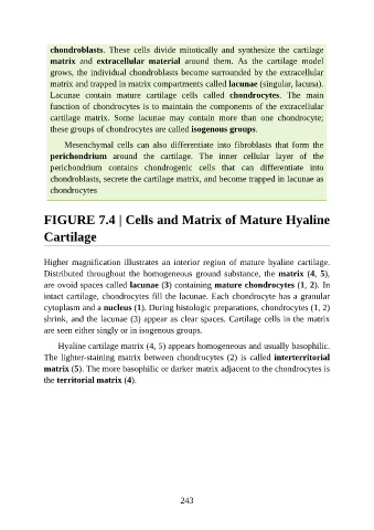
FIGURE 7.4 | Cells and Matrix of Mature Hyaline
Cartilage
Higher magnification illustrates an interior region of mature hyaline cartilage.
Distributed throughout the homogeneous ground substance, the matrix (4, 5),
are ovoid spaces called lacunae (3) containing mature chondrocytes (1, 2). In
intact cartilage, chondrocytes fill the lacunae. Each chondrocyte has a granular
cytoplasm and a nucleus (1). During histologic preparations, chondrocytes (1, 2)
shrink, and the lacunae (3) appear as clear spaces. Cartilage cells in the matrix
are seen either singly or in isogenous groups.
Hyaline cartilage matrix (4, 5) appears homogeneous and usually basophilic.
The lighter-staining matrix between chondrocytes (2) is called interterritorial
matrix (5). The more basophilic or darker matrix adjacent to the chondrocytes is
the territorial matrix (4).
243