
Page 245 - Atlas of Histology with Functional Correlations
P. 245
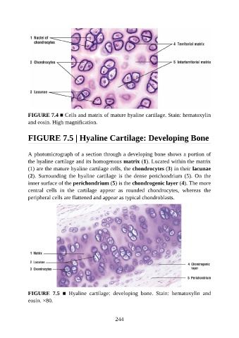
FIGURE 7.4 ■ Cells and matrix of mature hyaline cartilage. Stain: hematoxylin
               and eosin. High magnification.


               FIGURE 7.5 | Hyaline Cartilage: Developing Bone


               A photomicrograph of a section through a developing bone shows a portion of

               the hyaline cartilage and its homogenous matrix (1). Located within the matrix
               (1) are the mature hyaline cartilage cells, the chondrocytes (3) in their lacunae
               (2).  Surrounding  the  hyaline  cartilage  is  the  dense  perichondrium  (5).  On  the
               inner surface of the perichondrium (5) is the chondrogenic layer (4). The more

               central  cells  in  the  cartilage  appear  as  rounded  chondrocytes,  whereas  the
               peripheral cells are flattened and appear as typical chondroblasts.


               FIGURE  7.5  ■  Hyaline  cartilage:  developing  bone.  Stain:  hematoxylin  and
               eosin. ×80.


                                                          244