
Page 249 - Atlas of Histology with Functional Correlations
P. 249
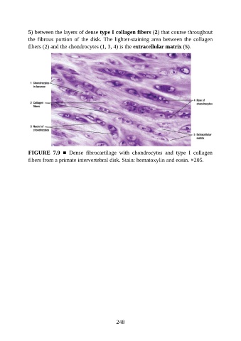
5) between the layers of dense type I collagen fibers (2) that course throughout

               the fibrous portion of the disk. The lighter-staining area between the collagen
               fibers (2) and the chondrocytes (1, 3, 4) is the extracellular matrix (5).


               FIGURE  7.9  ■  Dense  fibrocartilage  with  chondrocytes  and  type  I  collagen

               fibers from a primate intervertebral disk. Stain: hematoxylin and eosin. ×205.


                                                          248