
Page 491 - Atlas of Histology with Functional Correlations
P. 491
larger than eccrine sweat glands, and their ducts open into the hair follicle
canal. The secretory portion of the gland is coiled and tubular. However, in
contrast to eccrine sweat glands, the lumina of the secretory portion of the
gland are wide and dilated, and the secretory cells are low cuboidal. The
excretory ducts of the apocrine glands are also stratified cuboidal and are
similar to eccrine sweat glands. Similarly, the secretory portions of the
apocrine glands are surrounded by contractile myoepithelial cells. The
apocrine sweat glands become functional at puberty, when the sex hormones
are produced. The glands produce a viscous secretion, which acquires a
distinct and unpleasant odor after bacterial decomposition.
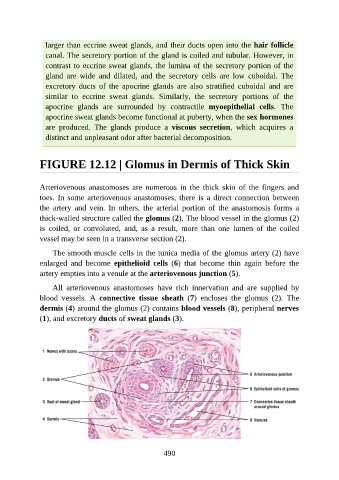
FIGURE 12.12 | Glomus in Dermis of Thick Skin
Arteriovenous anastomoses are numerous in the thick skin of the fingers and
toes. In some arteriovenous anastomoses, there is a direct connection between
the artery and vein. In others, the arterial portion of the anastomosis forms a
thick-walled structure called the glomus (2). The blood vessel in the glomus (2)
is coiled, or convoluted, and, as a result, more than one lumen of the coiled
vessel may be seen in a transverse section (2).
The smooth muscle cells in the tunica media of the glomus artery (2) have
enlarged and become epithelioid cells (6) that become thin again before the
artery empties into a venule at the arteriovenous junction (5).
All arteriovenous anastomoses have rich innervation and are supplied by
blood vessels. A connective tissue sheath (7) encloses the glomus (2). The
dermis (4) around the glomus (2) contains blood vessels (8), peripheral nerves
(1), and excretory ducts of sweat glands (3).
490