
Page 492 - Atlas of Histology with Functional Correlations
P. 492
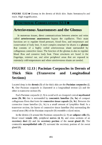
FIGURE 12.12 ■ Glomus in the dermis of thick skin. Stain: hematoxylin and

               eosin. High magnification.


                  FUNCTIONAL CORRELATIONS 12.3 ■


                 Arteriovenous Anastomoses and the Glomus


                     In numerous tissues, direct communications between arteries and veins
                 called  arteriovenous  anastomoses  bypass  the  capillaries.  Their  main

                 functions  are  to  regulate  blood  pressure,  blood  flow,  and  temperature  and
                 conservation of body heat. A more complex structure for shunts is a glomus
                 that  consists  of  a  highly  coiled  arteriovenous  shunt  surrounded  by
                 collagenous connective tissue. The function of the glomus is also to regulate

                 blood  flow  and  conserve  body  heat.  These  structures  are  found  in  the
                 fingertips,  external  ear,  and  other  peripheral  areas  that  are  exposed  to
                 extremely cold temperatures and where arteriovenous shunts are needed.


               FIGURE 12.13 | Pacinian Corpuscles in Dermis of

               Thick  Skin  (Transverse  and  Longitudinal


               Sections)


               Located deep in the dermis (3) of the thick skin are the Pacinian corpuscles (2,
               9).  One  Pacinian  corpuscle  is  illustrated  in  a  longitudinal  section  (2)  and  the
               other in transverse section (9).

                   Each Pacinian corpuscle (2, 9) is ovoid with an elongated central myelinated

               axon (2b, 9b) that is surrounded by concentric lamellae (2a, 9a) of compact
               collagenous fibers that form the connective tissue capsule (2c, 9c). Between the
               connective tissue lamellae (2c, 9c) is a small amount of lymphlike fluid. In a
               transverse section, the layers of connective tissue lamellae (9a) surrounding the

               central axon (9b) of the Pacinian corpuscle (9) resemble a sliced onion.

                   In the dermis (3) around the Pacinian corpuscles (2, 9) are adipose cells (5),
               blood  vessel  venule  (10),  peripheral  nerves  (4,  6),  and  cross  sections  of  an
               excretory  duct  (1)  and  the  secretory  portion  of  the  sweat  gland  (8).  The

               contractile myoepithelial cells (7) surround the secretory portion of the sweat
               gland (8).


                                                          491