
Page 488 - Atlas of Histology with Functional Correlations
P. 488
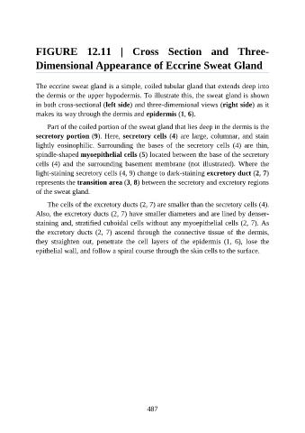
FIGURE  12.11  |  Cross  Section  and  Three-

               Dimensional Appearance of Eccrine Sweat Gland


               The eccrine sweat gland is a simple, coiled tubular gland that extends deep into
               the dermis or the upper hypodermis. To illustrate this, the sweat gland is shown

               in both cross-sectional (left side) and three-dimensional views (right side) as it
               makes its way through the dermis and epidermis (1, 6).

                   Part of the coiled portion of the sweat gland that lies deep in the dermis is the
               secretory portion (9). Here, secretory cells (4) are large, columnar, and stain

               lightly  eosinophilic.  Surrounding  the  bases  of  the  secretory  cells  (4)  are  thin,
               spindle-shaped myoepithelial cells (5) located between the base of the secretory
               cells (4) and the surrounding basement membrane (not illustrated). Where the
               light-staining secretory cells (4, 9) change to dark-staining excretory duct (2, 7)

               represents the transition area (3, 8) between the secretory and excretory regions
               of the sweat gland.

                   The cells of the excretory ducts (2, 7) are smaller than the secretory cells (4).
               Also, the excretory ducts (2, 7) have smaller diameters and are lined by denser-

               staining and, stratified cuboidal cells without any myoepithelial cells (2, 7). As
               the  excretory  ducts  (2,  7)  ascend  through  the  connective  tissue  of  the  dermis,
               they  straighten  out,  penetrate  the  cell  layers  of  the  epidermis  (1,  6),  lose  the
               epithelial wall, and follow a spiral course through the skin cells to the surface.


                                                          487