
Page 486 - Atlas of Histology with Functional Correlations
P. 486
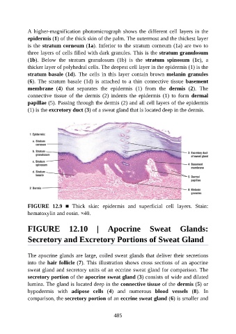
A higher-magnification photomicrograph shows the different cell layers in the

               epidermis (1) of the thick skin of the palm. The outermost and the thickest layer
               is the stratum corneum (1a). Inferior to the stratum corneum (1a) are two to
               three layers of cells filled with dark granules. This is the stratum granulosum
               (1b).  Below  the  stratum  granulosum  (1b)  is  the  stratum  spinosum  (1c),  a
               thicker layer of polyhedral cells. The deepest cell layer in the epidermis (1) is the

               stratum basale (1d). The cells in this layer contain brown melanin granules
               (6).  The  stratum  basale  (1d)  is  attached  to  a  thin  connective  tissue  basement
               membrane  (4)  that  separates  the  epidermis  (1)  from  the  dermis  (2).  The

               connective  tissue  of  the  dermis  (2)  indents  the  epidermis  (1)  to  form  dermal
               papillae (5). Passing through the dermis (2) and all cell layers of the epidermis
               (1) is the excretory duct (3) of a sweat gland that is located deep in the dermis.


               FIGURE  12.9  ■  Thick  skin:  epidermis  and  superficial  cell  layers.  Stain:
               hematoxylin and eosin. ×40.


               FIGURE  12.10  |  Apocrine  Sweat  Glands:

               Secretory and Excretory Portions of Sweat Gland


               The apocrine glands are large, coiled sweat glands that deliver their secretions
               into the hair follicle (7). This illustration shows cross sections of an apocrine
               sweat gland and secretory units of an eccrine sweat gland for comparison. The

               secretory portion of the apocrine sweat gland (3) consists of wide and dilated
               lumina. The gland is located deep in the connective tissue of the dermis (5) or
               hypodermis  with  adipose  cells  (4)  and  numerous  blood  vessels  (8).  In
               comparison, the secretory portion of an eccrine sweat gland (6) is smaller and


                                                          485