Page 485 - Atlas of Histology with Functional Correlations
P. 485
slide preparations. At a higher magnification, the outlines of flattened cells and

               eleidin droplets in this layer are occasionally seen.

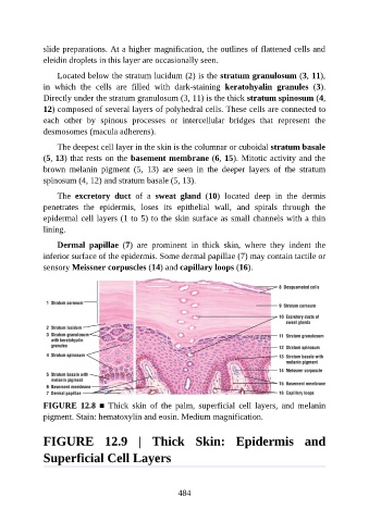
                   Located below the stratum lucidum (2) is the stratum granulosum (3, 11),
               in  which  the  cells  are  filled  with  dark-staining  keratohyalin  granules  (3).
               Directly under the stratum granulosum (3, 11) is the thick stratum spinosum (4,
               12) composed of several layers of polyhedral cells. These cells are connected to

               each  other  by  spinous  processes  or  intercellular  bridges  that  represent  the
               desmosomes (macula adherens).

                   The deepest cell layer in the skin is the columnar or cuboidal stratum basale
               (5, 13) that rests on the basement membrane (6, 15). Mitotic activity and the

               brown  melanin  pigment  (5,  13)  are  seen  in  the  deeper  layers  of  the  stratum
               spinosum (4, 12) and stratum basale (5, 13).

                   The  excretory  duct  of  a  sweat  gland  (10)  located  deep  in  the  dermis
               penetrates  the  epidermis,  loses  its  epithelial  wall,  and  spirals  through  the
               epidermal cell layers (1 to 5) to the skin surface as small channels with a thin

               lining.

                   Dermal  papillae  (7)  are  prominent  in  thick  skin,  where  they  indent  the
               inferior surface of the epidermis. Some dermal papillae (7) may contain tactile or
               sensory Meissner corpuscles (14) and capillary loops (16).


               FIGURE  12.8  ■  Thick  skin  of  the  palm,  superficial  cell  layers,  and  melanin
               pigment. Stain: hematoxylin and eosin. Medium magnification.


               FIGURE  12.9  |  Thick  Skin:  Epidermis  and

               Superficial Cell Layers


                                                          484

