
Page 484 - Atlas of Histology with Functional Correlations
P. 484
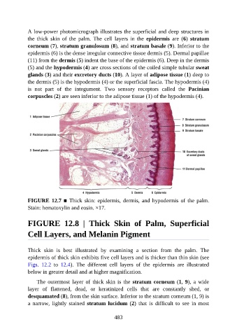
A low-power photomicrograph illustrates the superficial and deep structures in

               the  thick  skin  of  the  palm.  The  cell  layers  in  the  epidermis  are  (6)  stratum
               corneum (7), stratum granulosum (8), and stratum basale (9). Inferior to the
               epidermis (6) is the dense irregular connective tissue dermis (5). Dermal papillae
               (11) from the dermis (5) indent the base of the epidermis (6). Deep in the dermis
               (5) and the hypodermis (4) are cross sections of the coiled simple tubular sweat

               glands (3) and their excretory ducts (10). A layer of adipose tissue (1) deep to
               the dermis (5) is the hypodermis (4) or the superficial fascia. The hypodermis (4)
               is  not  part  of  the  integument.  Two  sensory  receptors  called  the  Pacinian

               corpuscles (2) are seen inferior to the adipose tissue (1) of the hypodermis (4).


               FIGURE 12.7 ■  Thick  skin:  epidermis,  dermis,  and  hypodermis  of  the  palm.
               Stain: hematoxylin and eosin. ×17.


               FIGURE  12.8  |  Thick  Skin  of  Palm,  Superficial

               Cell Layers, and Melanin Pigment


               Thick  skin  is  best  illustrated  by  examining  a  section  from  the  palm.  The
               epidermis of thick skin exhibits five cell layers and is thicker than thin skin (see
               Figs.  12.2  to  12.4).  The  different  cell  layers  of  the  epidermis  are  illustrated
               below in greater detail and at higher magnification.


                   The  outermost  layer  of  thick  skin  is  the  stratum corneum (1, 9),  a  wide
               layer  of  flattened,  dead,  or  keratinized  cells  that  are  constantly  shed,  or
               desquamated (8), from the skin surface. Inferior to the stratum corneum (1, 9) is
               a  narrow,  lightly  stained  stratum lucidum (2)  that  is  difficult  to  see  in  most


                                                          483