
Page 481 - Atlas of Histology with Functional Correlations
P. 481
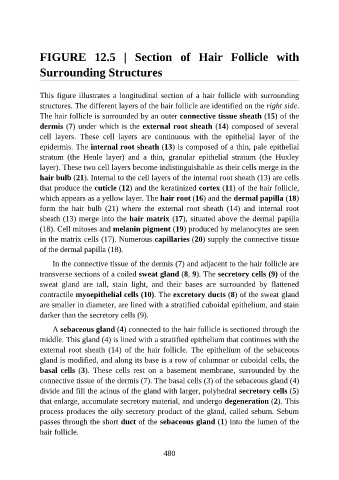
FIGURE  12.5  |  Section  of  Hair  Follicle  with

               Surrounding Structures


               This figure illustrates a longitudinal section of a hair follicle with surrounding
               structures. The different layers of the hair follicle are identified on the right side.

               The hair follicle is surrounded by an outer connective tissue sheath (15) of the
               dermis (7) under which is the external root sheath (14) composed of several
               cell  layers.  These  cell  layers  are  continuous  with  the  epithelial  layer  of  the

               epidermis. The internal root sheath (13) is composed of a thin, pale epithelial
               stratum  (the  Henle  layer)  and  a  thin,  granular  epithelial  stratum  (the  Huxley
               layer). These two cell layers become indistinguishable as their cells merge in the
               hair bulb (21). Internal to the cell layers of the internal root sheath (13) are cells
               that produce the cuticle (12) and the keratinized cortex (11) of the hair follicle,

               which appears as a yellow layer. The hair root (16) and the dermal papilla (18)
               form  the  hair  bulb  (21)  where  the  external  root  sheath  (14)  and  internal  root
               sheath (13) merge into the hair matrix (17), situated above the dermal papilla

               (18). Cell mitoses and melanin pigment (19) produced by melanocytes are seen
               in the matrix cells (17). Numerous capillaries (20) supply the connective tissue
               of the dermal papilla (18).

                   In the connective tissue of the dermis (7) and adjacent to the hair follicle are
               transverse sections of a coiled sweat gland (8, 9). The secretory cells (9) of the

               sweat  gland  are  tall,  stain  light,  and  their  bases  are  surrounded  by  flattened
               contractile myoepithelial cells (10). The excretory ducts (8) of the sweat gland
               are smaller in diameter, are lined with a stratified cuboidal epithelium, and stain
               darker than the secretory cells (9).


                   A sebaceous gland (4) connected to the hair follicle is sectioned through the
               middle. This gland (4) is lined with a stratified epithelium that continues with the
               external  root  sheath  (14)  of  the  hair  follicle.  The  epithelium  of  the  sebaceous
               gland is modified, and along its base is a row of columnar or cuboidal cells, the
               basal cells (3).  These  cells  rest  on  a  basement  membrane,  surrounded  by  the

               connective tissue of the dermis (7). The basal cells (3) of the sebaceous gland (4)
               divide and fill the acinus of the gland with larger, polyhedral secretory cells (5)
               that enlarge, accumulate secretory material, and undergo degeneration (2). This

               process produces the oily secretory product of the gland, called sebum. Sebum
               passes through the short duct of the sebaceous gland (1) into the lumen of the
               hair follicle.


                                                          480