
Page 477 - Atlas of Histology with Functional Correlations
P. 477
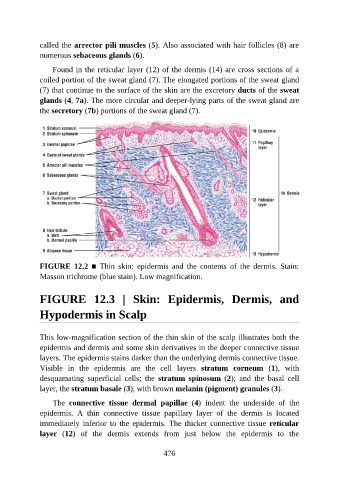
called the arrector pili muscles (5). Also associated with hair follicles (8) are

               numerous sebaceous glands (6).

                   Found in the reticular layer (12) of the dermis (14) are cross sections of a
               coiled portion of the sweat gland (7). The elongated portions of the sweat gland
               (7) that continue to the surface of the skin are the excretory ducts of the sweat
               glands (4, 7a). The more circular and deeper-lying parts of the sweat gland are

               the secretory (7b) portions of the sweat gland (7).


               FIGURE  12.2  ■  Thin  skin:  epidermis  and  the  contents  of  the  dermis.  Stain:
               Masson trichrome (blue stain). Low magnification.


               FIGURE  12.3  |  Skin:  Epidermis,  Dermis,  and

               Hypodermis in Scalp


               This low-magnification section of the thin skin of the scalp illustrates both the

               epidermis and dermis and some skin derivatives in the deeper connective tissue
               layers. The epidermis stains darker than the underlying dermis connective tissue.
               Visible  in  the  epidermis  are  the  cell  layers  stratum  corneum  (1),  with
               desquamating  superficial  cells;  the  stratum  spinosum  (2);  and  the  basal  cell
               layer, the stratum basale (3), with brown melanin (pigment) granules (3).


                   The  connective  tissue  dermal  papillae  (4)  indent  the  underside  of  the
               epidermis.  A  thin  connective  tissue  papillary  layer  of  the  dermis  is  located
               immediately  inferior  to  the  epidermis.  The  thicker  connective  tissue  reticular
               layer  (12)  of  the  dermis  extends  from  just  below  the  epidermis  to  the


                                                          476