
Page 478 - Atlas of Histology with Functional Correlations
P. 478
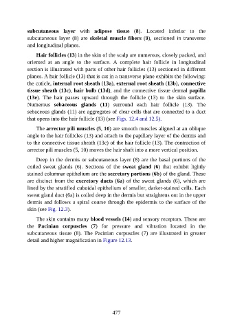
subcutaneous  layer  with  adipose  tissue  (8).  Located  inferior  to  the

               subcutaneous  layer  (8)  are  skeletal  muscle  fibers (9),  sectioned  in  transverse
               and longitudinal planes.

                   Hair follicles (13) in the skin of the scalp are numerous, closely packed, and
               oriented  at  an  angle  to  the  surface.  A  complete  hair  follicle  in  longitudinal
               section is illustrated with parts of other hair follicles (13) sectioned in different

               planes. A hair follicle (13) that is cut in a transverse plane exhibits the following:
               the cuticle, internal root sheath (13a), external root sheath (13b), connective
               tissue sheath (13c), hair bulb (13d), and the connective tissue dermal papilla
               (13e).  The  hair  passes  upward  through  the  follicle  (13)  to  the  skin  surface.

               Numerous  sebaceous  glands  (11)  surround  each  hair  follicle  (13).  The
               sebaceous glands (11) are aggregates of clear cells that are connected to a duct
               that opens into the hair follicle (13) (see Figs. 12.4 and 12.5).

                   The arrector pili muscles (5, 10) are smooth muscles aligned at an oblique
               angle to the hair follicles (13) and attach to the papillary layer of the dermis and

               to the connective tissue sheath (13c) of the hair follicle (13). The contraction of
               arrector pili muscles (5, 10) moves the hair shaft into a more vertical position.

                   Deep in the dermis or subcutaneous layer (8) are the basal portions of the
               coiled  sweat  glands  (6).  Sections  of  the  sweat  gland  (6)  that  exhibit  lightly

               stained columnar epithelium are the secretory portions (6b) of the gland. These
               are distinct from the excretory ducts (6a)  of  the  sweat  glands  (6),  which  are
               lined by the stratified cuboidal epithelium of smaller, darker-stained cells. Each
               sweat gland duct (6a) is coiled deep in the dermis but straightens out in the upper

               dermis and follows a spiral course through the epidermis to the surface of the
               skin (see Fig. 12.3).

                   The skin contains many blood vessels (14) and sensory receptors. These are
               the  Pacinian  corpuscles  (7)  for  pressure  and  vibration  located  in  the

               subcutaneous  tissue  (8).  The  Pacinian  corpuscles  (7)  are  illustrated  in  greater
               detail and higher magnification in Figure 12.13.


                                                          477