
Page 619 - Atlas of Histology with Functional Correlations
P. 619
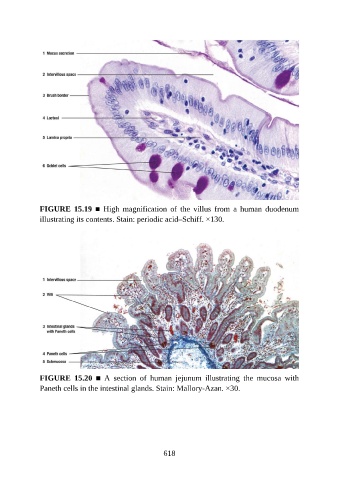
FIGURE  15.19  ■  High  magnification  of  the  villus  from  a  human  duodenum
               illustrating its contents. Stain: periodic acid–Schiff. ×130.


               FIGURE  15.20  ■  A  section  of  human  jejunum  illustrating  the  mucosa  with
               Paneth cells in the intestinal glands. Stain: Mallory-Azan. ×30.


                                                          618