
Page 620 - Atlas of Histology with Functional Correlations
P. 620
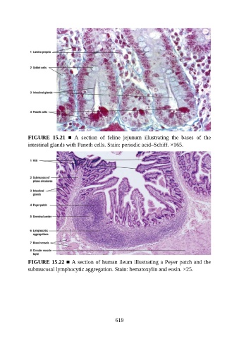
FIGURE  15.21  ■  A  section  of  feline  jejunum  illustrating  the  bases  of  the
               intestinal glands with Paneth cells. Stain: periodic acid–Schiff. ×165.


               FIGURE 15.22 ■ A section of human ileum illustrating a Peyer patch and the
               submucosal lymphocytic aggregation. Stain: hematoxylin and eosin. ×25.


                                                          619