
Page 624 - Atlas of Histology with Functional Correlations
P. 624
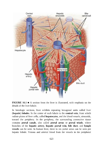
FIGURE 16.1 ■ A section from the liver is illustrated, with emphasis on the
               details of the liver lobule.

               In  histologic  sections,  liver  exhibits  repeating  hexagonal  units  called  liver
               (hepatic) lobules. In the center of each lobule is the central vein, from which

               radiate plates of liver cells, called hepatocytes, and the blood vessels, sinusoids,
               toward  the  periphery.  In  the  periphery,  the  surrounding  connective  tissue
               contains  portal  canals,  also  called  portal  areas  or  portal  triads,  where
               branches  of  the  hepatic  artery,  hepatic  portal  vein,  bile  duct,  and  lymph

               vessels can  be  seen.  In  human liver,  three  to  six  portal areas  can  be  seen  per
               hepatic  lobule.  Venous  and  arterial  blood  from  the  vessels  in  the  peripheral


                                                          623