
Page 626 - Atlas of Histology with Functional Correlations
P. 626
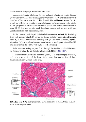
connective tissue septa (5, 9) that stain dark blue.

                   A complete hepatic lobule (on the left) and parts of adjacent hepatic lobules

               (7) are illustrated. The blue-staining interlobular septa (5, 9) contain interlobular
               branches of the portal vein (4, 11), bile duct (2, 12), and hepatic artery (3, 13),
               which are collectively considered as portal areas, portal canals, or portal triads.
               At the periphery of each lobule are several portal areas within the interlobular

               septa  (5,  9)  that  also  contain  small  lymphatic  vessels  and  nerves,  which  are
               usually small and only occasionally seen.

                   In the center of each hepatic lobule (7) is the central vein (1, 8). Radiating
               from each central vein (1, 8) toward the lobule periphery are plates of hepatic

               cells  (6).  Located  between  the  hepatic  plates  (6)  are  blood  channels,  hepatic
               sinusoids (10). Arterial  and  venous  blood  mixes in  the  hepatic  sinusoids  (10)
               and flows toward the central vein (1, 8) of each lobule (7).

                   Bile, produced by hepatocytes, flows through the tiny bile canaliculi between
               the hepatocytes into the interlobular bile ducts (2, 12) (see Fig. 16.6).

                   The interlobular vessels and bile ducts (2 to 4, 11 to 13) are highly branched,

               and,  in  a  cross  section  of  the  liver  lobule,  more  than  one  section  of  these
               structures are seen within a portal area.


               FIGURE 16.2 ■ Pig liver (panoramic view, transverse section). Stain: Mallory-
               Azan. Low magnification.


                                                          625