
Page 627 - Atlas of Histology with Functional Correlations
P. 627
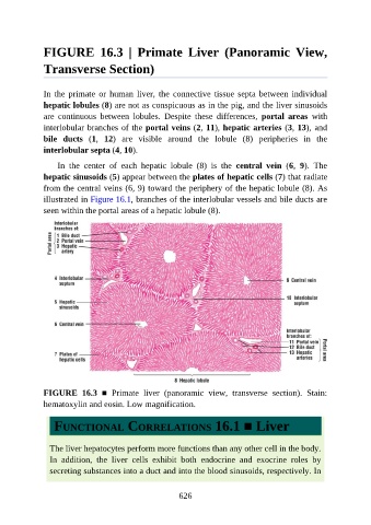
FIGURE  16.3  |  Primate  Liver  (Panoramic  View,

               Transverse Section)


               In  the  primate  or  human  liver,  the  connective  tissue  septa  between  individual
               hepatic lobules (8) are not as conspicuous as in the pig, and the liver sinusoids

               are  continuous  between  lobules.  Despite  these  differences,  portal  areas  with
               interlobular branches of the portal veins (2, 11), hepatic arteries (3, 13), and
               bile  ducts  (1,  12)  are  visible  around  the  lobule  (8)  peripheries  in  the
               interlobular septa (4, 10).

                   In  the  center  of  each  hepatic  lobule  (8)  is  the  central  vein  (6,  9).  The

               hepatic sinusoids (5) appear between the plates of hepatic cells (7) that radiate
               from the central veins (6, 9) toward the periphery of the hepatic lobule (8). As
               illustrated in Figure 16.1, branches of the interlobular vessels and bile ducts are

               seen within the portal areas of a hepatic lobule (8).


               FIGURE  16.3  ■  Primate  liver  (panoramic  view,  transverse  section).  Stain:
               hematoxylin and eosin. Low magnification.


                  FUNCTIONAL CORRELATIONS 16.1 ■ Liver


                 The liver hepatocytes perform more functions than any other cell in the body.

                 In  addition,  the  liver  cells  exhibit  both  endocrine  and  exocrine  roles  by
                 secreting substances into a duct and into the blood sinusoids, respectively. In


                                                          626