
Page 630 - Atlas of Histology with Functional Correlations
P. 630
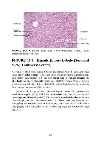
FIGURE  16.4  ■  Bovine  liver:  liver  lobule  (transverse  section).  Stain:
               hematoxylin and eosin. ×30.


               FIGURE 16.5 | Hepatic (Liver) Lobule (Sectional

               View, Transverse Section)


               A  section  of  the  hepatic  lobule  between  the  central  vein  (9)  and  connective
               tissue interlobular septum (1, 6) of the portal area is illustrated in greater detail.

               In the interlobular septum (1, 6) are seen portal vein (4), hepatic arteries (3),
               bile ducts (5),  and  a  lymphatic vessel (2).  Multiple  cross  sections  of  hepatic
               arteries (3) and bile ducts (5) are attributable to their branching in the septum or
               their passage into and out of the septum.

                   Branches  of  the  portal  vein  (4)  and  hepatic  artery  (3)  penetrate  the

               interlobular  septum  (1,  6)  and  form  the  sinusoids  (8,  10)  that  are  situated
               between plates of hepatic cells (7). Discontinuous endothelial cells (10) line the
               sinusoids  (8,  10)  and  the  central  vein  (9).  Blood  cells  (erythrocytes  and

               leukocytes)  in  sinusoids (8)  drain  toward  the  central  vein  (9)  of  each  lobule.
               Also present in the sinusoids (10) are fixed macrophages, the Kupffer cells (see
               Fig. 16.7).


                                                          629