
Page 633 - Atlas of Histology with Functional Correlations
P. 633
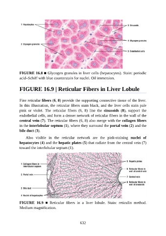
FIGURE 16.8 ■ Glycogen granules in liver cells (hepatocytes). Stain: periodic
               acid–Schiff with blue counterstain for nuclei. Oil immersion.


               FIGURE 16.9 | Reticular Fibers in Liver Lobule


               Fine reticular fibers (6, 8) provide the supporting connective tissue of the liver.
               In this illustration, the reticular fibers stain black, and the liver cells stain pale

               pink  or  violet.  The  reticular  fibers  (6,  8)  line  the  sinusoids  (8),  support  the
               endothelial cells, and form a denser network of reticular fibers in the wall of the
               central vein (7). The reticular fibers (6, 8) also merge with the collagen fibers
               in the interlobular septum (1), where they surround the portal vein (2) and the

               bile duct (3).

                   Also  visible  in  the  reticular  network  are  the  pink-staining  nuclei  of
               hepatocytes (4) and the hepatic plates (5) that radiate from the central vein (7)
               toward the interlobular septum (1).


               FIGURE  16.9  ■  Reticular  fibers  in  a  liver  lobule.  Stain:  reticulin  method.
               Medium magnification.


                                                          632