
Page 638 - Atlas of Histology with Functional Correlations
P. 638
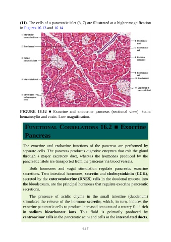
(11). The cells of a pancreatic islet (3, 7) are illustrated at a higher magnification

               in Figures 16.13 and 16.14.


               FIGURE  16.12  ■  Exocrine  and  endocrine  pancreas  (sectional  view).  Stain:
               hematoxylin and eosin. Low magnification.


                  FUNCTIONAL CORRELATIONS 16.2 ■ Exocrine


                  Pancreas


                 The  exocrine  and  endocrine  functions  of  the  pancreas  are  performed  by
                 separate cells. The pancreas produces digestive enzymes that exit the gland
                 through  a  major  excretory  duct,  whereas  the  hormones  produced  by  the

                 pancreatic islets are transported from the pancreas via blood vessels.

                     Both  hormones  and  vagal  stimulation  regulate  pancreatic  exocrine
                 secretions. Two intestinal hormones, secretin and cholecystokinin (CCK),
                 secreted by the enteroendocrine (DNES) cells in the duodenal mucosa into
                 the bloodstream, are the principal hormones that regulate exocrine pancreatic

                 secretions.

                     The  presence  of  acidic  chyme  in  the  small  intestine  (duodenum)
                 stimulates the release of the hormone secretin, which, in turn, induces the
                 exocrine pancreatic cells to produce increased amounts of a watery fluid rich

                 in  sodium  bicarbonate  ions.  This  fluid  is  primarily  produced  by
                 centroacinar cells in the pancreatic acini and cells in the intercalated ducts.


                                                          637