
Page 643 - Atlas of Histology with Functional Correlations
P. 643
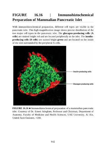
FIGURE                  16.16             |        Immunohistochemical

               Preparation of Mammalian Pancreatic Islet


               With  immunohistochemical  preparation,  different  cell  types  are  visible  in  the
               pancreatic islet. This high-magnification image shows precise distribution of the

               two major cell types in the pancreatic islet. The glucagon-producing cells (A
               cells) are stained bright red and are located peripherally in the islet. The insulin-
               producing cells (B cells) are stained bright green and are located on the inside
               of the islet surrounded by the peripheral A cells.


               FIGURE 16.16 ■ Immunohistochemical preparation of a mammalian pancreatic

               islet. Courtesy of Dr. Ernest Adeghate, Professor and Chairman, Department of
               Anatomy, Faculty of Medicine and Health Sciences, UAE University, Al Ain,
               United Arab Emirates. ×200.


                                                          642