
Page 640 - Atlas of Histology with Functional Correlations
P. 640
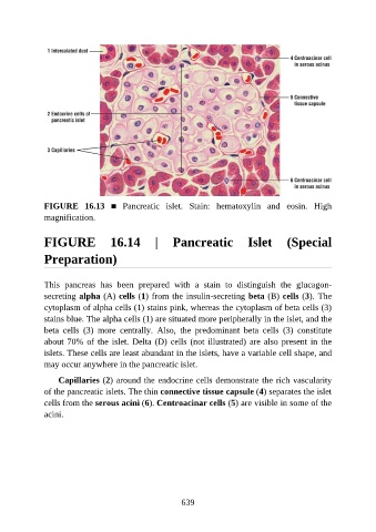
FIGURE  16.13  ■  Pancreatic  islet.  Stain:  hematoxylin  and  eosin.  High
               magnification.


               FIGURE  16.14  |  Pancreatic  Islet  (Special

               Preparation)


               This  pancreas  has  been  prepared  with  a  stain  to  distinguish  the  glucagon-
               secreting alpha (A) cells (1) from the insulin-secreting beta (B) cells (3). The
               cytoplasm of alpha cells (1) stains pink, whereas the cytoplasm of beta cells (3)
               stains blue. The alpha cells (1) are situated more peripherally in the islet, and the

               beta  cells  (3)  more  centrally.  Also,  the  predominant  beta  cells  (3)  constitute
               about 70% of the islet. Delta (D) cells (not illustrated) are also present in the
               islets. These cells are least abundant in the islets, have a variable cell shape, and
               may occur anywhere in the pancreatic islet.

                   Capillaries (2) around the endocrine cells demonstrate the rich vascularity

               of the pancreatic islets. The thin connective tissue capsule (4) separates the islet
               cells from the serous acini (6). Centroacinar cells (5) are visible in some of the
               acini.


                                                          639