
Page 637 - Atlas of Histology with Functional Correlations
P. 637
cells. Each group of these cells secretes a single hormone.
Alpha cells constitute about 20% of the islets and are located around the islet
periphery. The most numerous beta cells constitute about 70% of the islet cells
and are concentrated in the center of the islet. The remaining cell types are few
in number and are located throughout the islets.
Supplemental micrographic images are available at
www.thePoint.com/Eroschenko13e under Digestive System Part IV: Liver,
Pancreas, and Gallbladder.
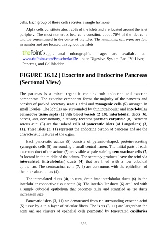
FIGURE 16.12 | Exocrine and Endocrine Pancreas
(Sectional View)
The pancreas is a mixed organ; it contains both endocrine and exocrine
components. The exocrine component forms the majority of the pancreas and
consists of packed secretory serous acini and zymogenic cells (5) arranged in
small lobules. The lobules are surrounded by thin intralobular and interlobular
connective tissue septa (1) with blood vessels (2, 10), interlobular ducts (6),
nerves, and, occasionally, a sensory receptor pacinian corpuscle (8). Between
serous acini (5) are the isolated cells of pancreatic islets (of Langerhans) (3,
11). These islets (3, 11) represent the endocrine portion of pancreas and are the
characteristic features of the organ.
Each pancreatic acinus (5) consists of pyramid-shaped, protein-secreting
zymogenic cells (5) surrounding a small central lumen. The initial parts of each
excretory duct of the acinus (5) are visible as pale-staining centroacinar cells (7,
9) located in the middle of the acinus. The secretory products leave the acini via
intercalated (intralobular) ducts (4) that are lined with a low cuboidal
epithelium. The centroacinar cells (7, 9) are continuous with the epithelium of
the intercalated ducts (4).
The intercalated ducts (4), in turn, drain into interlobular ducts (6) in the
interlobular connective tissue septa (4). The interlobular ducts (6) are lined with
a simple cuboidal epithelium that becomes taller and stratified as the ducts
increase in size.
Pancreatic islets (3, 11) are demarcated from the surrounding exocrine acini
(5) tissue by a thin layer of reticular fibers. The islets (3, 11) are larger than the
acini and are clusters of epithelial cells permeated by fenestrated capillaries
636