
Page 639 - Atlas of Histology with Functional Correlations
P. 639
The bicarbonate fluid neutralizes the acidic chyme, stops the action of the
proteolytic enzyme pepsin secreted by gastric glands in the stomach, and
creates a neutral environment in the duodenum for the digestive pancreatic
enzymes.
The presence of fats and proteins in the small intestine induces CCK
release that stimulates the pancreatic acinar cells to secrete digestive
enzymes. These are pancreatic amylase for carbohydrate digestion,
pancreatic lipase for lipid digestion, deoxyribonuclease and ribonuclease
for digestion of nucleic acids, and the proteolytic enzymes trypsinogen,
chymotrypsinogen, and procarboxypeptidase.
Pancreatic enzymes are produced in the acinar cells, released in an
inactive form after hormonal stimulation, and are indirectly activated only in
the lumen of the duodenum by the hormone enterokinase secreted by the
intestinal mucosa. Enterokinase, in turn, converts trypsinogen to trypsin, and
trypsin then converts all inactive pancreatic enzymes into active digestive
enzymes in the chyme.
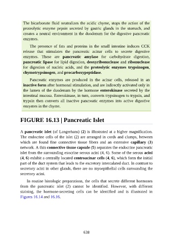
FIGURE 16.13 | Pancreatic Islet
A pancreatic islet (of Langerhans) (2) is illustrated at a higher magnification.
The endocrine cells of the islet (2) are arranged in cords and clumps, between
which are found fine connective tissue fibers and an extensive capillary (3)
network. A thin connective tissue capsule (5) separates the endocrine pancreatic
islet from the surrounding exocrine serous acini (4, 6). Some of the serous acini
(4, 6) exhibit a centrally located centroacinar cells (4, 6), which form the initial
part of the duct system that leads to the excretory intercalated duct. In contrast to
secretory acini in other glands, there are no myoepithelial cells surrounding the
secretory acini.
In routine histologic preparations, the cells that secrete different hormones
from the pancreatic islet (2) cannot be identified. However, with different
staining, the hormone-secreting cells can be identified and is illustrated in
Figures 16.14 and 16.16.
638