
Page 644 - Atlas of Histology with Functional Correlations
P. 644
SECTION 3 Gallbladder
The gallbladder is a small, hollow organ attached to the inferior surface of the
liver. Bile is produced by liver hepatocytes that leaves the liver and flows to, is
stored, and is concentrated in the gallbladder. Upon hormonal stimulation, bile
leaves the gallbladder via the cystic duct and enters the duodenum via the
common bile duct through the major duodenal papilla, a finger-like protrusion
of the duodenal wall into the lumen.
The gallbladder is not a gland. Its main function is to store and concentrate
bile by absorbing its water. Bile is released into the digestive tract as a result of
hormonal stimulation after a meal that contains fatty foods. When the gallbladder
is empty, the mucosa exhibits deep folds.
Supplemental micrographic images are available at
www.thePoint.com/Eroschenko13e under Digestive System Part IV: Liver,
Pancreas, and Gallbladder.
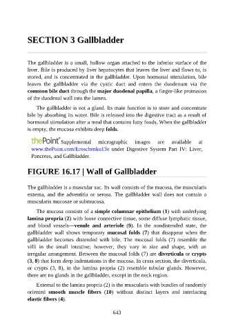
FIGURE 16.17 | Wall of Gallbladder
The gallbladder is a muscular sac. Its wall consists of the mucosa, the muscularis
externa, and the adventitia or serosa. The gallbladder wall does not contain a
muscularis mucosae or submucosa.
The mucosa consists of a simple columnar epithelium (1) with underlying
lamina propria (2) with loose connective tissue, some diffuse lymphatic tissue,
and blood vessels—venule and arteriole (9). In the nondistended state, the
gallbladder wall shows temporary mucosal folds (7) that disappear when the
gallbladder becomes distended with bile. The mucosal folds (7) resemble the
villi in the small intestine; however, they vary in size and shape, with an
irregular arrangement. Between the mucosal folds (7) are diverticula or crypts
(3, 8) that form deep indentations in the mucosa. In cross section, the diverticula,
or crypts (3, 8), in the lamina propria (2) resemble tubular glands. However,
there are no glands in the gallbladder, except in the neck region.
External to the lamina propria (2) is the muscularis with bundles of randomly
oriented smooth muscle fibers (10) without distinct layers and interlacing
elastic fibers (4).
643