
Page 645 - Atlas of Histology with Functional Correlations
P. 645
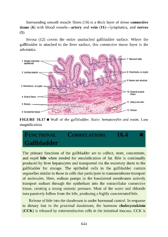
Surrounding smooth muscle fibers (10) is a thick layer of dense connective

               tissue (6)  with  blood  vessels—artery  and  vein (11)—lymphatics,  and  nerves
               (5).

                   Serosa  (12)  covers  the  entire  unattached  gallbladder  surface.  Where  the
               gallbladder  is  attached  to  the  liver  surface,  this  connective  tissue  layer  is  the
               adventitia.


               FIGURE 16.17 ■ Wall of the gallbladder. Stain: hematoxylin and eosin. Low

               magnification.


                  FUNCTIONAL                     CORRELATIONS                       16.4            ■

                  Gallbladder


                 The  primary  functions  of  the  gallbladder  are  to  collect,  store,  concentrate,

                 and  expel  bile  when  needed  for  emulsification  of  fat.  Bile  is  continually
                 produced by liver hepatocytes and transported via the excretory ducts to the
                 gallbladder  for  storage.  The  epithelial  cells  in  the  gallbladder  contain
                 organelles similar to those in cells that participate in transmembrane transport
                 of  molecules.  Here,  sodium  pumps  in  the  basolateral  membranes  actively

                 transport  sodium  through  the  epithelium  into  the  extracellular  connective
                 tissue,  creating  a  strong  osmotic  pressure.  Most  of  the  water  and  chloride
                 ions passively follow from the bile, producing a highly concentrated bile.

                     Release of bile into the duodenum is under hormonal control. In response

                 to  dietary  fats  in  the  proximal  duodenum,  the  hormone  cholecystokinin
                 (CCK) is released by enteroendocrine cells in the intestinal mucosa. CCK is


                                                          644