
Page 634 - Atlas of Histology with Functional Correlations
P. 634
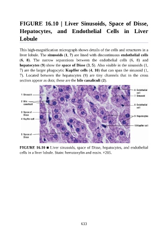
FIGURE  16.10  |  Liver  Sinusoids,  Space  of  Disse,

               Hepatocytes,  and  Endothelial  Cells  in  Liver

               Lobule


               This high-magnification micrograph shows details of the cells and structures in a
               liver lobule. The sinusoids (1, 7) are lined with discontinuous endothelial cells
               (6,  8).  The  narrow  separations  between  the  endothelial  cells  (6,  8)  and
               hepatocytes (9) show the space of Disse (3, 5). Also visible in the sinusoids (1,
               7) are the larger phagocytic Kupffer cells (4, 10) that can span the sinusoid (1,

               7).  Located  between  the  hepatocytes  (9)  are  tiny  channels  that  in  the  cross
               section appear as dots; these are the bile canaliculi (2).


               FIGURE 16.10 ■ Liver sinusoids, space of Disse, hepatocytes, and endothelial
               cells in a liver lobule. Stain: hematoxylin and eosin. ×205.


                                                          633