
Page 631 - Atlas of Histology with Functional Correlations
P. 631
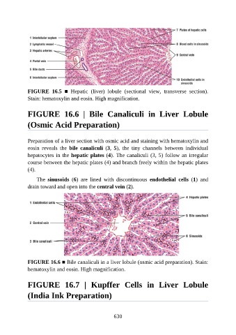
FIGURE  16.5  ■  Hepatic  (liver)  lobule  (sectional  view,  transverse  section).
               Stain: hematoxylin and eosin. High magnification.


               FIGURE  16.6  |  Bile  Canaliculi  in  Liver  Lobule

               (Osmic Acid Preparation)


               Preparation of a liver section with osmic acid and staining with hematoxylin and
               eosin  reveals  the  bile  canaliculi  (3,  5),  the  tiny  channels  between  individual
               hepatocytes in the hepatic plates (4). The canaliculi (3, 5) follow an irregular
               course between the hepatic plates (4) and branch freely within the hepatic plates

               (4).

                   The  sinusoids  (6)  are  lined  with  discontinuous  endothelial  cells  (1)  and
               drain toward and open into the central vein (2).


               FIGURE 16.6 ■ Bile canaliculi in a liver lobule (osmic acid preparation). Stain:
               hematoxylin and eosin. High magnification.


               FIGURE  16.7  |  Kupffer  Cells  in  Liver  Lobule


               (India Ink Preparation)


                                                          630