
Page 632 - Atlas of Histology with Functional Correlations
P. 632
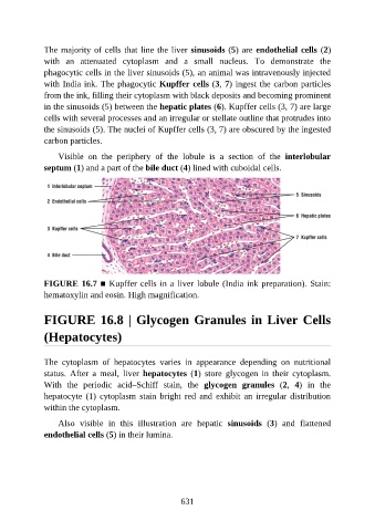
The majority of cells that line the liver sinusoids (5) are endothelial cells (2)

               with  an  attenuated  cytoplasm  and  a  small  nucleus.  To  demonstrate  the
               phagocytic cells in the liver sinusoids (5), an animal was intravenously injected
               with India ink. The phagocytic Kupffer cells (3, 7) ingest the carbon particles
               from the ink, filling their cytoplasm with black deposits and becoming prominent
               in the sinusoids (5) between the hepatic plates (6). Kupffer cells (3, 7) are large

               cells with several processes and an irregular or stellate outline that protrudes into
               the sinusoids (5). The nuclei of Kupffer cells (3, 7) are obscured by the ingested
               carbon particles.

                   Visible  on  the  periphery  of  the  lobule  is  a  section  of  the  interlobular

               septum (1) and a part of the bile duct (4) lined with cuboidal cells.


               FIGURE 16.7 ■ Kupffer cells in a liver lobule (India ink preparation). Stain:
               hematoxylin and eosin. High magnification.


               FIGURE 16.8 | Glycogen Granules in Liver Cells


               (Hepatocytes)


               The  cytoplasm  of  hepatocytes  varies  in  appearance  depending  on  nutritional
               status.  After  a  meal,  liver  hepatocytes  (1)  store  glycogen  in  their  cytoplasm.

               With  the  periodic  acid–Schiff  stain,  the  glycogen  granules  (2,  4)  in  the
               hepatocyte  (1)  cytoplasm  stain  bright  red  and  exhibit  an  irregular  distribution
               within the cytoplasm.

                   Also  visible  in  this  illustration  are  hepatic  sinusoids  (3)  and  flattened

               endothelial cells (5) in their lumina.


                                                          631