
Page 655 - Atlas of Histology with Functional Correlations
P. 655
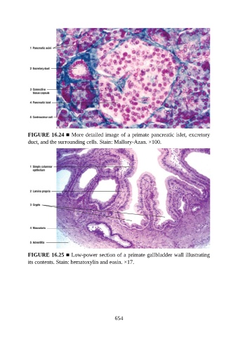
FIGURE 16.24 ■ More detailed image of a primate pancreatic islet, excretory

               duct, and the surrounding cells. Stain: Mallory-Azan. ×100.


               FIGURE 16.25 ■ Low-power section of a primate gallbladder wall illustrating
               its contents. Stain: hematoxylin and eosin. ×17.


                                                          654