
Page 657 - Atlas of Histology with Functional Correlations
P. 657
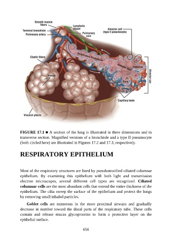
FIGURE 17.1 ■ A section of the lung is illustrated in three dimensions and in
               transverse section. Magnified versions of a bronchiole and a type II pneumocyte
               (both circled here) are illustrated in Figures 17.2 and 17.3, respectively.


               RESPIRATORY EPITHELIUM


               Most of the respiratory structures are lined by pseudostratified ciliated columnar

               epithelium.  By  examining  this  epithelium  with  both  light  and  transmission
               electron  microscopes,  several  different  cell  types  are  recognized.  Ciliated
               columnar cells are the most abundant cells that extend the entire thickness of the
               epithelium. The cilia sweep the surface of the epithelium and protect the lungs
               by removing small inhaled particles.


                   Goblet  cells  are  numerous  in  the  more  proximal  airways  and  gradually
               decrease in number toward the distal parts of the respiratory tube. These cells
               contain  and  release  mucus  glycoproteins  to  form  a  protective  layer  on  the
               epithelial surface.


                                                          656