
Page 661 - Atlas of Histology with Functional Correlations
P. 661
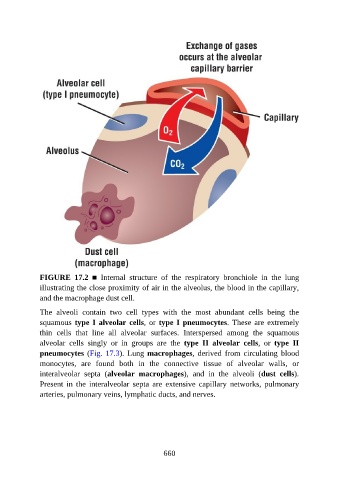
FIGURE  17.2  ■  Internal  structure  of  the  respiratory  bronchiole  in  the  lung
               illustrating the close proximity of air in the alveolus, the blood in the capillary,

               and the macrophage dust cell.

               The  alveoli  contain  two  cell  types  with  the  most  abundant  cells  being  the
               squamous type I alveolar cells, or type I pneumocytes. These are extremely
               thin  cells  that  line  all  alveolar  surfaces.  Interspersed  among  the  squamous
               alveolar  cells  singly  or  in  groups  are  the  type  II  alveolar  cells,  or  type  II

               pneumocytes (Fig. 17.3). Lung macrophages,  derived  from  circulating  blood
               monocytes,  are  found  both  in  the  connective  tissue  of  alveolar  walls,  or
               interalveolar  septa  (alveolar  macrophages),  and  in  the  alveoli  (dust  cells).

               Present  in  the  interalveolar  septa  are  extensive  capillary  networks,  pulmonary
               arteries, pulmonary veins, lymphatic ducts, and nerves.


                                                          660