
Page 663 - Atlas of Histology with Functional Correlations
P. 663
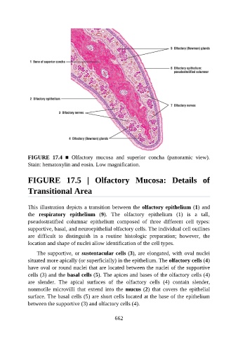
FIGURE  17.4  ■  Olfactory  mucosa  and  superior  concha  (panoramic  view).

               Stain: hematoxylin and eosin. Low magnification.


               FIGURE  17.5  |  Olfactory  Mucosa:  Details  of


               Transitional Area


               This illustration depicts a transition between the olfactory epithelium (1) and
               the  respiratory  epithelium  (9).  The  olfactory  epithelium  (1)  is  a  tall,
               pseudostratified  columnar  epithelium  composed  of  three  different  cell  types:

               supportive, basal, and neuroepithelial olfactory cells. The individual cell outlines
               are  difficult  to  distinguish  in  a  routine  histologic  preparation;  however,  the
               location and shape of nuclei allow identification of the cell types.

                   The supportive, or sustentacular cells (3), are elongated, with oval nuclei

               situated more apically (or superficially) in the epithelium. The olfactory cells (4)
               have oval or round nuclei that are located between the nuclei of the supportive
               cells (3) and the basal cells (5). The apices and bases of the olfactory cells (4)
               are  slender.  The  apical  surfaces  of  the  olfactory  cells  (4)  contain  slender,

               nonmotile  microvilli  that  extend  into  the  mucus (2)  that  covers  the  epithelial
               surface. The basal cells (5) are short cells located at the base of the epithelium
               between the supportive (3) and olfactory cells (4).


                                                          662