
Page 665 - Atlas of Histology with Functional Correlations
P. 665
photomicrograph.
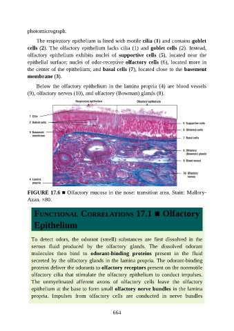
The respiratory epithelium is lined with motile cilia (1) and contains goblet
cells (2). The olfactory epithelium lacks cilia (1) and goblet cells (2). Instead,
olfactory epithelium exhibits nuclei of supportive cells (5), located near the
epithelial surface; nuclei of odor-receptive olfactory cells (6), located more in
the center of the epithelium; and basal cells (7), located close to the basement
membrane (3).
Below the olfactory epithelium in the lamina propria (4) are blood vessels
(9), olfactory nerves (10), and olfactory (Bowman) glands (8).
FIGURE 17.6 ■ Olfactory mucosa in the nose: transition area. Stain: Mallory-
Azan. ×80.
FUNCTIONAL CORRELATIONS 17.1 ■ Olfactory
Epithelium
To detect odors, the odorant (smell) substances are first dissolved in the
serous fluid produced by the olfactory glands. The dissolved odorant
molecules then bind to odorant-binding proteins present in the fluid
secreted by the olfactory glands in the lamina propria. The odorant-binding
proteins deliver the odorants to olfactory receptors present on the nonmotile
olfactory cilia that stimulate the olfactory epithelium to conduct impulses.
The unmyelinated afferent axons of olfactory cells leave the olfactory
epithelium at the base to form small olfactory nerve bundles in the lamina
propria. Impulses from olfactory cells are conducted in nerve bundles
664