
Page 667 - Atlas of Histology with Functional Correlations
P. 667
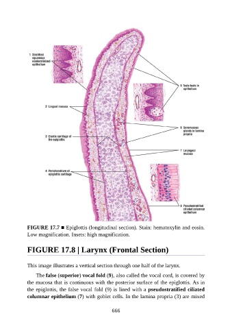
FIGURE 17.7 ■ Epiglottis (longitudinal section). Stain: hematoxylin and eosin.
               Low magnification. Insets: high magnification.


               FIGURE 17.8 | Larynx (Frontal Section)


               This image illustrates a vertical section through one half of the larynx.

                   The false (superior) vocal fold (9), also called the vocal cord, is covered by

               the mucosa that is continuous with the posterior surface of the epiglottis. As in
               the epiglottis, the false vocal fold (9) is lined with a pseudostratified ciliated
               columnar epithelium (7) with goblet cells. In the lamina propria (3) are mixed


                                                          666