
Page 670 - Atlas of Histology with Functional Correlations
P. 670
the mucosa. Most of the trachealis muscle (7) fibers insert into the
perichondrium (9) that covers the hyaline cartilage (3).
The lumen of the trachea is lined with a pseudostratified ciliated columnar
epithelium (12) with goblet cells. The underlying lamina propria (13) contains
connective tissue fibers, diffuse lymphatic tissue, and occasional lymphatic
nodules. Located deeper in the lamina propria (13) is the longitudinal elastic
membrane (14) formed by elastic fibers. This membrane (14) divides the lamina
propria (13) from the submucosa (4) in which are found the tubuloacinar
seromucous tracheal glands (10) whose excretory ducts (11) pass through the
lamina propria (13) to the tracheal lumen.
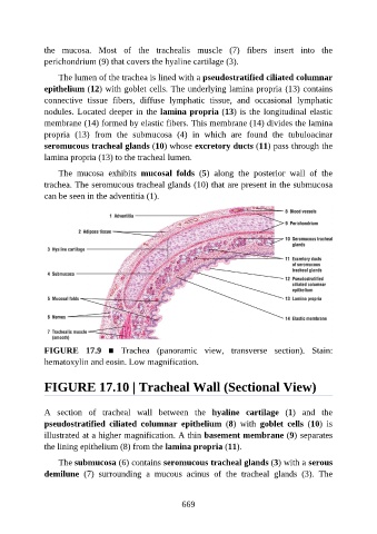
The mucosa exhibits mucosal folds (5) along the posterior wall of the
trachea. The seromucous tracheal glands (10) that are present in the submucosa
can be seen in the adventitia (1).
FIGURE 17.9 ■ Trachea (panoramic view, transverse section). Stain:
hematoxylin and eosin. Low magnification.
FIGURE 17.10 | Tracheal Wall (Sectional View)
A section of tracheal wall between the hyaline cartilage (1) and the
pseudostratified ciliated columnar epithelium (8) with goblet cells (10) is
illustrated at a higher magnification. A thin basement membrane (9) separates
the lining epithelium (8) from the lamina propria (11).
The submucosa (6) contains seromucous tracheal glands (3) with a serous
demilune (7) surrounding a mucous acinus of the tracheal glands (3). The
669