
Page 674 - Atlas of Histology with Functional Correlations
P. 674
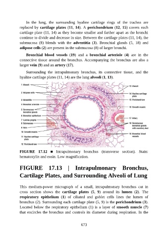
In  the  lung,  the  surrounding  hyaline  cartilage  rings  of  the  trachea  are

               replaced by cartilage plates (11, 14). A perichondrium (12, 15) covers each
               cartilage plate (11, 14) as they become smaller and farther apart as the bronchi
               continue to divide and decrease in size. Between the cartilage plates (11, 14), the
               submucosa  (8)  blends  with  the  adventitia  (3).  Bronchial  glands  (5,  18)  and
               adipose cells (2) are present in the submucosa (8) of larger bronchi.


                   Bronchial  blood  vessels  (19)  and  a  bronchial  arteriole  (4)  are  in  the
               connective tissue around the bronchus. Accompanying the bronchus are also a
               larger vein (9) and an artery (17).

                   Surrounding  the  intrapulmonary  bronchus,  its  connective  tissue,  and  the

               hyaline cartilage plates (11, 14) are the lung alveoli (1, 13).


               FIGURE  17.12  ■  Intrapulmonary  bronchus  (transverse  section).  Stain:

               hematoxylin and eosin. Low magnification.


               FIGURE  17.13  |  Intrapulmonary  Bronchus,


               Cartilage Plates, and Surrounding Alveoli of Lung


               This  medium-power  micrograph  of  a  small,  intrapulmonary  bronchus  cut  in
               cross  section  shows  the  cartilage  plates  (5,  9)  around  its  lumen  (2).  The
               respiratory  epithelium  (1)  of  ciliated  and  goblet  cells  lines  the  lumen  of

               bronchus (2). Surrounding each cartilage plate (5, 9) is the perichondrium (3).
               Located below the respiratory epithelium (1) is a layer of smooth muscle (7)
               that encircles the bronchus and controls its diameter during respiration. In the


                                                          673