
Page 675 - Atlas of Histology with Functional Correlations
P. 675
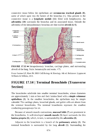
connective  tissue  below  the  epithelium  are  seromucous  tracheal  glands  (8),

               some  of  which  open  into  the  lumen  of  the  bronchus  (1).  Also  present  in  the
               connective  tissue  is  a  lymphatic  nodule  (11)  filled  with  lymphocytes.  An
               adventitia  (10)  surrounds  the  bronchus  and  its  associated  tissue.  Outside  the
               adventitia of the intrapulmonary bronchus are thin-walled alveoli (4, 6).


               FIGURE  17.13  ■  Intrapulmonary  bronchus,  cartilage  plates,  and  surrounding
               alveoli of the lung. Stain: hematoxylin and eosin. ×75.

               From: Gartner LP, Hiatt JM. BRS Cell Biology & Histology. 6th ed. Baltimore: Lippincott
               Williams & Wilkins, 2011.


               FIGURE 17.14 | Terminal Bronchiole (Transverse

               Section)


               The  bronchioles  subdivide  into  smaller  terminal  bronchioles,  whose  diameters

               are approximately 1 mm or less and their lumina lined with a simple columnar
               epithelium  (3).  In  the  smallest  bronchioles,  the  epithelium  may  be  simple
               cuboidal. The cartilage plates, bronchial glands, and goblet cells are absent from

               the  terminal  bronchioles.  The  terminal  bronchioles  represent  the  smallest
               conducting passageways for air.

                   Because of smooth muscle contractions, mucosal folds (7) are prominent in
               the bronchioles. A well-developed smooth muscle (5) layer surrounds the thin
               lamina propria (6), which, in turn, is surrounded by the adventitia (8).

                   Adjacent  to  the  bronchiole  is  a  branch  of  the  pulmonary artery (2). The

               terminal  bronchiole  is  surrounded  by  the  lung  alveoli  (1).  Surrounding  the


                                                          674