
Page 673 - Atlas of Histology with Functional Correlations
P. 673
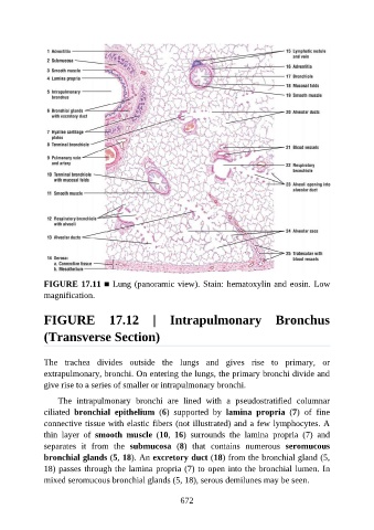
FIGURE 17.11 ■ Lung (panoramic view). Stain: hematoxylin and eosin. Low
               magnification.


               FIGURE  17.12  |  Intrapulmonary  Bronchus

               (Transverse Section)


               The  trachea  divides  outside  the  lungs  and  gives  rise  to  primary,  or

               extrapulmonary, bronchi. On entering the lungs, the primary bronchi divide and
               give rise to a series of smaller or intrapulmonary bronchi.

                   The  intrapulmonary  bronchi  are  lined  with  a  pseudostratified  columnar
               ciliated  bronchial  epithelium  (6)  supported  by  lamina  propria  (7)  of  fine
               connective tissue with elastic fibers (not illustrated) and a few lymphocytes. A

               thin  layer  of  smooth  muscle  (10,  16)  surrounds  the  lamina  propria  (7)  and
               separates  it  from  the  submucosa  (8)  that  contains  numerous  seromucous
               bronchial glands (5, 18). An excretory duct (18) from the bronchial gland (5,
               18) passes through the lamina propria (7) to open into the bronchial lumen. In

               mixed seromucous bronchial glands (5, 18), serous demilunes may be seen.


                                                          672