
Page 676 - Atlas of Histology with Functional Correlations
P. 676
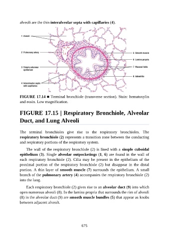
alveoli are the thin interalveolar septa with capillaries (4).


               FIGURE 17.14 ■ Terminal bronchiole (transverse section). Stain: hematoxylin
               and eosin. Low magnification.


               FIGURE 17.15 | Respiratory Bronchiole, Alveolar

               Duct, and Lung Alveoli


               The  terminal  bronchioles  give  rise  to  the  respiratory  bronchioles.  The
               respiratory bronchiole (2) represents a transition zone between the conducting
               and respiratory portions of the respiratory system.

                   The wall of the respiratory bronchiole (2) is lined with a simple cuboidal
               epithelium (3). Single alveolar outpocketings (1, 6) are found in the wall of

               each  respiratory  bronchiole  (2).  Cilia  may  be  present  in  the  epithelium  of  the
               proximal  portion  of  the  respiratory  bronchiole  (2)  but  disappear  in  the  distal
               portion. A thin layer of smooth muscle (7) surrounds the epithelium. A small
               branch of the pulmonary artery (4) accompanies the respiratory bronchiole (2)

               into the lung.

                   Each respiratory bronchiole (2) gives rise to an alveolar duct (9) into which
               open numerous alveoli (8). In the lamina propria that surrounds the rim of alveoli
               (8) in the alveolar duct (9) are smooth muscle bundles (5) that appear as knobs
               between adjacent alveoli.


                                                          675