
Page 680 - Atlas of Histology with Functional Correlations
P. 680
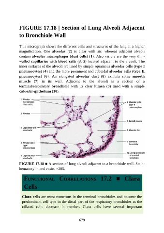
FIGURE 17.18 | Section of Lung Alveoli Adjacent

               to Bronchiole Wall


               This micrograph shows the different cells and structures of the lung at a higher
               magnification.  One  alveolus  (2)  is  clear  with  air,  whereas  adjacent  alveoli

               contain alveolar macrophages (dust cells) (1). Also visible are the very thin-
               walled capillaries with blood cells (3, 5) located adjacent to the alveoli. The
               inner surfaces of the alveoli are lined by simple squamous alveolar cells (type I

               pneumocytes) (4) and the more prominent and cuboidal alveolar cells (type II
               pneumocytes)  (6).  An  elongated  alveolar  duct  (8)  exhibits  some  smooth
               muscle  (7)  in  its  wall.  Adjacent  to  the  alveoli  is  a  section  of  a
               terminal/respiratory  bronchiole  with  its  clear  lumen  (9)  lined  with  a  simple
               cuboidal epithelium (10).


               FIGURE 17.18 ■ A section of lung alveoli adjacent to a bronchiole wall. Stain:

               hematoxylin and eosin. ×205.


                  FUNCTIONAL  CORRELATIONS  17.2  ■  Clara

                  Cells


                 Clara cells are most numerous in the terminal bronchioles and become the
                 predominant cell type in the distal part of the respiratory bronchioles as the

                 ciliated  cells  decrease  in  number.  Clara  cells  have  several  important


                                                          679