
Page 682 - Atlas of Histology with Functional Correlations
P. 682
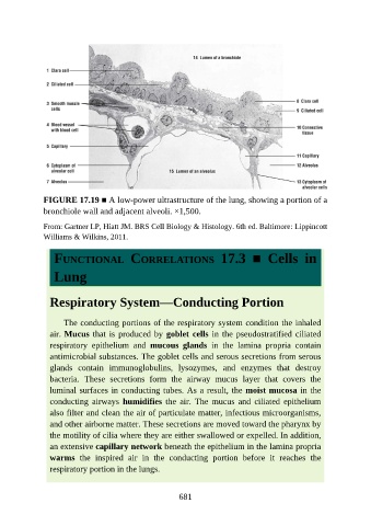
FIGURE 17.19 ■ A low-power ultrastructure of the lung, showing a portion of a

               bronchiole wall and adjacent alveoli. ×1,500.

               From: Gartner LP, Hiatt JM. BRS Cell Biology & Histology. 6th ed. Baltimore: Lippincott
               Williams & Wilkins, 2011.


                  FUNCTIONAL  CORRELATIONS  17.3  ■  Cells  in

                  Lung


                 Respiratory System—Conducting Portion


                     The conducting portions of the respiratory system condition the inhaled
                 air. Mucus that is produced by goblet cells in the pseudostratified ciliated
                 respiratory  epithelium  and  mucous  glands  in  the  lamina  propria  contain

                 antimicrobial substances. The goblet cells and serous secretions from serous
                 glands  contain  immunoglobulins,  lysozymes,  and  enzymes  that  destroy
                 bacteria.  These  secretions  form  the  airway  mucus  layer  that  covers  the
                 luminal surfaces in conducting tubes. As a result, the moist mucosa in the

                 conducting  airways  humidifies  the  air.  The  mucus  and  ciliated  epithelium
                 also filter and clean the air of particulate matter, infectious microorganisms,
                 and other airborne matter. These secretions are moved toward the pharynx by
                 the motility of cilia where they are either swallowed or expelled. In addition,

                 an extensive capillary network beneath the epithelium in the lamina propria
                 warms  the  inspired  air  in  the  conducting  portion  before  it  reaches  the
                 respiratory portion in the lungs.


                                                          681