
Page 678 - Atlas of Histology with Functional Correlations
P. 678
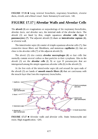
FIGURE  17.16  ■  Lung:  terminal  bronchiole,  respiratory  bronchiole,  alveolar

               ducts, alveoli, and a blood vessel. Stain: hematoxylin and eosin. ×40.


               FIGURE 17.17 | Alveolar Walls and Alveolar Cells


               The alveoli (3) are evaginations or outpocketings of the respiratory bronchioles,

               alveolar ducts, and alveolar sacs, the terminal ends of the alveolar ducts. The
               alveoli  (3)  are  lined  by  thin,  simple  squamous  alveolar  cells  (type  I
               pneumocytes) (7). The adjacent alveoli (3) share an interalveolar septum (4),
               or alveolar wall.


                   The interalveolar septa (4) consist of simple squamous alveolar cells (7), fine
               connective  tissue  fibers  and  fibroblasts,  and  numerous  capillaries (1)  that  are
               close to the alveolar cells (7) of the adjacent alveoli (3).

                   The  alveoli  (3)  also  contain  alveolar  macrophages  (6)  or  dust  cells  that
               normally contain several carbon or dust particles in their cytoplasm. Also in the

               alveoli  (3)  are  the  alveolar  cells  (2,  5)  or  type  II  pneumocytes  that  are
               interspersed among the simple squamous alveolar cells (6) in the alveoli (3).

                   At the free ends of the interalveolar septa (4) and around the open ends of
               the alveoli (3) are bands of smooth muscle fibers (8) that are continuous with

               the muscle layer that lines the respiratory bronchioles.


               FIGURE  17.17  ■  Alveolar  walls  and  alveolar  cells.  Stain:  hematoxylin  and
               eosin. High magnification. ×205.


                                                          677